Absence Crises or Common Distractions: What's the Difference?

Many of the clients with whom we intervene in our clinic face difficulties related to maintaining attention on a task, with quality and for satisfactory periods of time. Intervention with recourse to Neurofeedbackwith these people it has a high success rate, especially in cases where the so-called attention difficulties are associated with the excessive presence of low frequency rhythms (or, if we like, “slow activity”) in the electroencephalogram of the user. These low-frequency rhythms may be present constantly and continuously, or they may occur for periods of short seconds, with greater or lesser intensity.
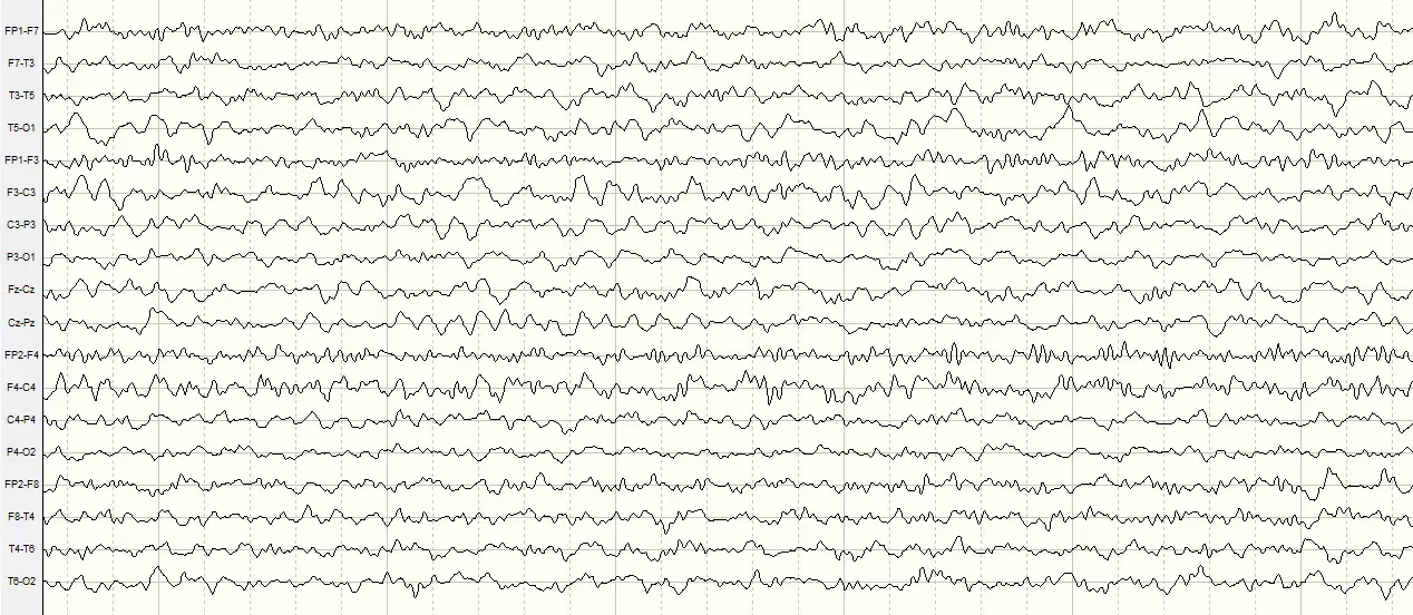
In most cases of diagnoses of Attention Deficit, it is possible to observe the excessive and relatively constant presence of slow rhythms, as exemplified by the brain map presented in Figure 1, below, compared to a Normal Brain Standard:

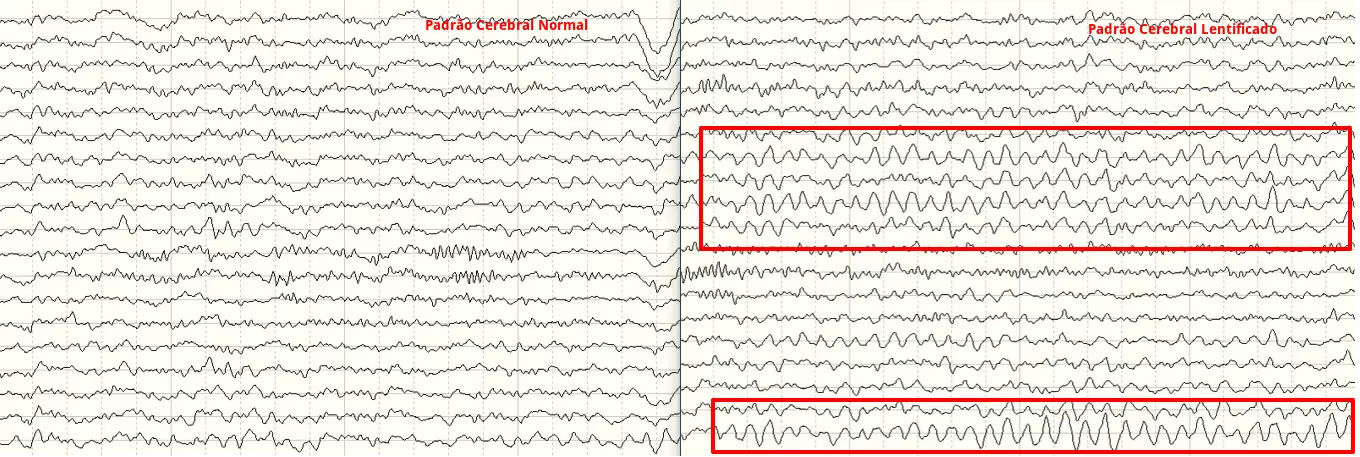
However, in many cases we find the presence of slow activity sporadically, present for brief seconds and which, as a consequence of this brevity, is less appreciable in the brain maps resulting from the electroencephalogram. In fact, these people can present a brain map very close to ideal in the lower frequency bands, but have serious difficulties in maintaining attention due to the presence of these sporadic “peaks” of slow activity.
Figure 2 shows on the left the plot of an electroencephalogram in which the pattern of brain activity is normal and on the right the plot of an electroencephalogram in which this type of activity is evident, and is indicated by the red rectangle:

It is visible, by comparison with the remaining EEG trace, that the areas marked by the red rectangle have a different morphology, with a greater amplitude, more “rounded” and with a lower frequency. These periods correspond to the aforementioned “peaks” of slow activity, periods that are strongly related to periods in which the attention of the person in question deviates from the task at hand, loses focus and resumes after a few seconds.
This description corresponds to a common distraction. At these times, the person is easily “called back” to the task they were performing and does not manifest other symptoms beyond the aforementioned loss of focus.
On the other hand, absence seizures are manifestations of certain epilepsy patterns. Its manifestation presents symptoms very similar to a simple and common distraction and, for this reason, they are often devalued without the diagnosis being made immediately.
Absence crises are more common in children between the ages of 4 and 14, and are misinterpreted as lack of attention and distraction. They are often associated with learning difficulties.
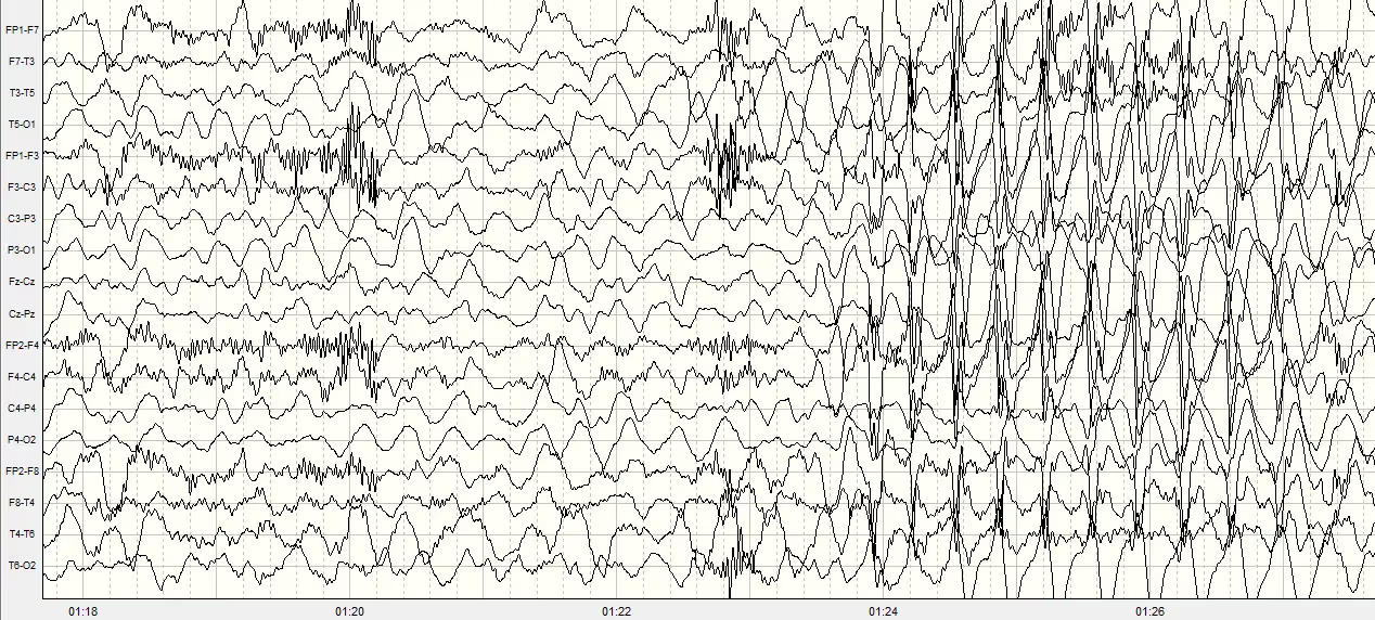
Figure 3 shows the changes in EEG during an absence crisis compared to a normal situation. It is possible to see a generalized slowdown of the pattern, with greater amplitude in the bilateral frontal region, always associated and interspersed with more abrupt activity with a bee-shaped format, which is characteristic of this type of epilepsy.


Figure 3
During an absence crisis, which can last between 10 to 20 seconds, we can observe that the child abruptly stops the activity he is doing, his gaze is stopped/fixed, sometimes some muscular or motor changes may be visible, such as constant blinking at an abnormally fast frequency, lip bursting or chewing movements and/or; movements with the hands.
During this brief period in which the child is having a seizure, it does not react to any sound, visual or tactile stimuli, and when the crisis ends the child can resume the activity as if nothing had happened or may suffer a brief period of confusion, always without memory of what happened.
In this way, and when there is a suspicion of Epilepsy, it is necessary that the people around the child, such as parents, relatives, caregivers and teachers/educators are attentive and know how to interpret and know the differences between a normal distraction from an absence crisis, so that the correct diagnosis and intervention are made as quickly as possible.
In summary and summary, the main differences are the following:
Common Distraction:
- It happens more easily in moments/situations that leave the child bored;
- It starts slowly;
- It may be interrupted;
- It tends to continue after a stimulus.
Absence Crisis:
- It can occur in any situation, including during the practice of physical activity;
- It starts suddenly, without any warning;
- It cannot be interrupted;
- Ends in about 10-20 seconds.
We Train Brains, Strengthen Minds, Transform Lives
Schedule an appointment and see how we can help your family.
No time for a call now? Leave your details and we will get in touch:

Wonderful team, concerned, attentive and always available to help in everything.
With the passage of time, the results are being verified.
I am grateful to have met Neuroimprove and all its professionals.


